These methods are :
1) X-ray Crystallography
2) Electron Microscopy (EM)
- Transmission EM
- Scanning EM
- STEM
3) Ultracentrifugation
- Purification
Now, I shall talk about the first physical method, which is X-ray Crystallography
X-RAY CRYSTALLOGRAPHY
 Source : http://www.district87.org/staff/sutterm/X-Ray/X-Ray/Intro%20to%20Xray%20Crys.htm
 What is X -Ray Crystallography?
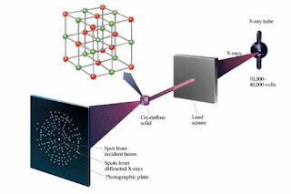
It is the study of crystal structures by applying the x-ray diffraction techniques.
Basically, when an Xray beam bombards a crystalline lattice in a specific type of orientation, the X-ray beam is divided in a definite way that was characterised by the lattice's atomic structure. This phenomenon is known as X-Ray diffraction and occurs when the wavelength of X-rays and the interatomic distances within the lattice have the exact magnitude order.
There are three main steps in X-ray crystallography.
1st Step (Often the most challenging step) :
Obtain an adequate crystal of material under study. The crystal should be large enough, maybe larger than 100 micrometres in all dimensions, pure in composition and has a regular structure, with no significant internal imperfections (cracks/twinning).
Conversely, a small or irregular crystal will give fewer and less accurate data, whereby it may be impossible to determine the atomic arrangement.
2nd step:
The crystal is placed in an intense beam of X-rays, usually monochromatic X-rays, that produces the regular pattern of reflections. Previous reflections disappear and new ones appear as the crystal is gradually rotated. The intensity of each spot is recorded at each single orientation of the crystal. In this case, several data sets may have to be collected with each set covering slightly more than half a complete rotation of the crystal and usually containing ten thousands of reflection intensities.
3rd step :
These collected data are compiled computationally with complementary chemical information in order to create and refine a model of the atomic arrangement within the crystal itself. The refined and final model of the atomic arrangement (Crystal Structure) is usually stored in a public database.
ELECTRON MICROSCOPY
What is an electron microscope?
An electron microscope is a type of microscope that uses electrons to illuminate a specimen and create an enlarged image. Electron microscopes have much greater resolving power than light microscopes and can obtain much higher magnifications.
ADD EM FROM PICTURES.
Transmission electron microscope:
The original form of electron microscope, the transmission electron microscope (TEM) uses a high voltage electron beam to create an image. The electrons are emitted by an electron gun, commonly fitted with a tungsten filament cathode as the electron source.
Scanning electron microscope:
Unlike the TEM, where electrons of the high voltage beam carry the image of the specimen, the electron beam of the Scanning Electron Microscope (SEM) does not at any time carry a complete image of the specimen. The SEM produces images by probing the specimen with a focused electron beam that is scanned across a rectangular area of the specimen.
Reflection electron microscope:
In the Reflection Electron Microscope (REM) as in the TEM, an electron beam is incident on a surface, but instead of using the transmission (TEM) or secondary electrons (SEM), the reflected beam of elastically scattered electrons is detected. This technique is typically coupled with Reflection High Energy Electron Diffraction and Reflection high-energy loss spectrum (RHELS).
Scanning Transmission Electron Microscope (STEM):
The STEM rasters a focused incident probe across a specimen that (as with the TEM) has been thinned to facilitate detection of electrons scattered through the specimen. The high resolution of the TEM is thus possible in STEM. The focusing action (and aberrations) occur before the electrons hit the specimen in the STEM, but afterward in the TEM.
ULTRACENTRIFUGATION
What is ultracentrifugation?
Ultracentrifugation is a centrifuge optimized for spinning a rotor at very high speeds, capable of generating acceleration as high as 1,000,000 g (9,800 km/s²). There are two kinds of ultracentrifuges, the preparative and the analytical ultracentrifuge.
PUT ULTRACENTRIFUDGE 1 IN PICTURES.
Source : http://www.mmb.usyd.edu.au/facilities/images/analytical_ultracentrifugation300x211.jpg
No comments:
Post a Comment