1) Analysis of viral proteins
. PAGE/ SDS PAGE
. Western blot
. Protein Sequencing
. X-ray crystallography
2) Analysis of viral genome
. Agarose gels
. Restriction analysis
. Sequencing
. Southern blot
. Northern blot
. PCR/R-T PCR
Now, allow me to explain the first part of the first main point, which is SDS PAGE.
SDS PAGE(PolyAcrylamide Gel Electrophoresis)
SDS
Since we are trying to separate many different protein molecules of different shapes and sizes, we first want to denatured so that the proteins no longer have any secondary, tertiary or quaternary structure (i.e. we want them to retain only their primary amino acid structure). We use SDS to denature all proteins to the same linear shape.
The pink line represent protein.
Source :http://www.bio.davidson.edu/courses/genomics/method/SDSPAGE/SDSPAGE.htmlSDS (sodium dodecyl sulfate) is a detergent (soap) that can dissolve hydrophobic molecules but also has a negative charge (sulfATE) attached to it. Therefore, if a cell is incubated with SDS, the membranes will be dissolved, all the proteins will be soluablized by the detergent, plus all the proteins will be covered with many negative charges. So a protein that started out like the one shown in the top part of figure will be converted into the one shown in the bottom part of figure.
The end result has two important features:
1) all proteins retain only their primary structure
2) all proteins have a large negative charge which means they will all migrate towards the positve pole when placed in an electric field.
PAGE
If the proteins are denatured and put into an electric field, they will all move towards the positive pole at the same rate, with no separation by size. So we need to put the proteins into an environment that will allow different sized proteins to move at different rates. The environment of choice is polyacrylamide, which is a polymer of acrylamide monomers. When this polymer is formed, it turns into a gel and we will use electricity to pull the proteins through the gel so the entire process is called polyacrylamide gel electrophoresis (PAGE). A polyacrylamide gel is not solid but is made of a laberynth of tunnels through a meshwork of fibers.
 Figure 2. This cartoon shows a slab of polyacrylamide (dark gray) with tunnels (different sized red rings with shading to depict depth) exposed on the edge. Notice that there are many different sizes of tunnels scattered randomly throughout the gel.
Now we are ready to apply the mixture of denatured proteins to the gel and apply the current. If all the proteins enter the gel at the same time and have the same force pulling them towards the other end, which ones will be able to move through the gel faster? Because of their small size, small molecules can manuver through the polyacrylamide forest faster than big molecules.Figure 3. This is a top view of two selected tunnels (only two are shown for clarity of the diagram). These tunnels extend all the way through the gel, but they meander through the gel and do not go in straight lines. Notice the difference in diameter of the two tunnels.
 Figure 4. Cartoon showing a mixutre of denatured proteins (pink lines of differen lengths) beginning their journey through a polyacrylamide gel (gray slab with tunnels). An electric filed is established with the positive pole (red plus) at the far end and the negative pole (black minus) at the closer end. Since all the proteins have strong negative charges, they will all move in the direction the arrow is pointing (run to red).
 When running an SDS-PAGE, we never let the proteins electrophorese (run) so long that they actually reach the other side of the gel. We turn off the current and then stain the proteins and see how far they moved through the gel (until we stain them, they are colorless and thus invisible). Notice that the actual bands are equal in size, but the proteins within each band are of different sizes.

WESTERN BLOT
Western blotting allows the detection of specific proteins from extracts made from cells or tissues, before or after any purification steps. Proteins are generally separated by size using gel electrophoresis before being transferred to a synthetic membrane via dry, semi-dry, or wet blotting methods.
Western blotting is a routine molecular biology method that can be used to semi-quantitatively compare protein levels between extracts.

http://search.msn.com.sg/images/results.aspx?q=western+blot&FORM=MSNH#
PROTEIN SEQUENCING
Proteins are found in every cell and are essential to every biological process, protein structure is very complex: determining a protein's structure involves first protein sequencing - determining the amino acid sequences of its constituent peptides; and also determining what conformation it adopts and whether it is complexed with any non-peptide molecules. Discovering the structures and functions of proteins in living organisms is an important tool for understanding cellular processes, and allows drugs that target specific metabolic pathways to be invented more easily.
X-RAY CRYSTALLOGRAPHY
X-ray crystallography is a method of determining the arrangement of atoms within a crystal, in which a beam of X-rays strikes a crystal and scatters into many different directions. From the angles and intensities of these scattered beams, a crystallographer can produce a three-dimensional picture of the density of electrons within the crystal.
The method revealed the structure and functioning of many biological molecules, including vitamins, drugs, proteins and nucleic acids such as DNA.
Now, I shall talk about the second main point (Analysis of viral genome)
AGAROSE GELS
Agarose gel electrophoresis is a method used in biochemistry and molecular biology to separate DNA, or RNA molecules by size. This is achieved by moving negatively charged nucleic acid molecules through an agarose matrix with an electric field (electrophoresis). Shorter molecules move faster and migrate farther than longer ones.

http://search.live.com/images/results.aspx?q=agarose+gels&FORM=BIRE#
RESTRICTION ANALYSIS
Restriction Analysis tools find restriction sites in nucleotide sequences using over 1000 enzymes from a restriction enzyme database, usually REBASE.
Reaction enzymes are proteins produced by bacteria that cleave DNA at specific sites along the molecule. In the bacterial cell, restriction enzymes cleave foreign DNA, thus eliminating infecting organisms. Restriction enzymes can be isolated from bacterial cells and used in the laboratory to manipulate fragments of DNA, such as those that contain genes; for this reason they are indispensable tools of recombinant DNA technology.
Each restriction enzyme recognizes a short, specific sequence of nucleotide bases. When a restriction endonuclease recognizes a sequence, it snips through the DNA molecule by catalyzing the hydrolysis (splitting of a chemical bond by addition of a water molecule) of the bond between adjacent nucleotides.

http://search.msn.com.sg/images/results.aspx?q=restriction+analysis&FORM=MSNH#
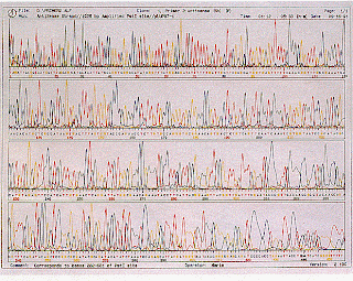
DNA SEQUENCING
DNA sequencing, the process of determining the exact order of the 3 billion chemical building blocks (called bases and abbreviated A, T, C, and G) that make up the DNA of the 24 different human chromosomes, was the greatest technical challenge in the Human Genome Project. Achieving this goal has helped reveal the estimated 20,000-25,000 human genes within our DNA as well as the regions controlling them. The resulting DNA sequence maps are being used by 21st Century scientists to explore human biology and other complex phenomena.
DNA SEQUENCING
http://www.youtube.com/watch?v=ezAefHhvecM
SEQUENCING (protein)
Protein sequencing provides the amino acid sequence at the N-terminus of your sample. It can be used to positively identify your protein and peptide.
Proteins are found in every cell and are essential to every biological process, protein structure is very complex: determining a protein's structure involves first protein sequencing - determining the amino acid sequences of its constituent peptides; and also determining what conformation it adopts and whether it is complexed with any non-peptide molecules. Discovering the structures and functions of proteins in living organisms is an important tool for understanding cellular processes, and allows drugs that target specific metabolic pathways to be invented more easily.
The two major direct methods of protein sequencing are mass spectrometry and the Edman degradation reaction. It is also possible to generate an amino acid sequence from the DNA or mRNA sequence encoding the protein, if this is known. However, there are a number of other reactions which can be used to gain more limited information about protein sequences and can be used as preliminaries to the aforementioned methods of sequencing or to overcome specific inadequacies within them.
SOUTHERN BLOT
The Southern blot method for marking specific DNA sequences and detecting the presence of a specific gene. The Southern blot is used for transferring DNA. After electrophoresis, the gel is treated with an alkaline to cause the DNA to denature and separate into single strands. A membranous sheet is placed on the gel, and pressure applied evenly via suction or the mundane method of paper towels with a weight. The DNA migrates to the membrane and sticks there. The DNA-impregnanted membrane is baked or radiated to permanently attach the DNA. Molecules are next treated with a hybridization probe, which is simply a DNA molecule with a known sequence that will pair with the blotted DNA's sequences. The probe DNA is tagged with fluorescent or chromogenic dyes so it can be identified.
NORTHERN BLOT
The Northern blot is used to analyze RNA. Northern blots can be used to determine the size of an mRNA transcript, identify functional variants of a gene and measure the relatedness of species. In the Northern blot, a blotting membrane is applied to a gel electrophoreses to get the molecules to transfer from the gel to the membrane. The gel is treated with a formaldehyde hybridization buffer to denature the RNA. The membrane should also be kept moist throughout. After pressure is applied evenly to the membrane, the RNA migrates and sticks to the membrane. Then the membrane is baked or irradiated, permanently attaching the RNA to the membrane. Northern blots are intended to study gene expression through identification and examination of genetic differences.

http://search.live.com/images/results.aspx?q=northern++blot&FORM=AWIR#\
POLYMERASE CHAIN REACTION (PCR)
PCR/RT-PCR
How PCR works. http://www.bio.davidson.edu/Courses/immunology/Flash/RT_PCR.html
Reverse transcription polymerase chain reaction, abbreviated as RT-PCR, is a laboratory technique for amplifying a defined piece of a ribonucleic acid (RNA) molecule. The RNA strand is first reverse transcribed into its DNA complement or complementary DNA, followed by amplification of the resulting DNA using polymerase chain reaction. This can either be a 1 or 2 step process. Polymerase chain reaction (PCR) itself is the process used to amplify specific parts of a DNA molecule, via the temperature-mediated enzyme DNA polymerase.
The two-step RT-PCR process for converting RNA to DNA subsequent PCR amplification of the reversely-transcribed DNA:
1. first strand reaction
Complementary DNA (cDNA) is made from an mRNA template using dNTPs & reverse transcriptase. The components are combined with a DNA primer in a reverse transcriptase buffer for an hour at 37°C.
2. second strand reaction


 http://www.ideascientific.com/images/south_blot.gif