Let's move on to the detection, identification and diagnosis of viruses. okay one of the tissue culture methods detection is PLAQUE ASSAY. What is it and how does it work? Let's find out together, shall we?
Plaque Assay:
A method of quantifying the number of infectious units by inoculating serial dilutions of a viral suspension on a cell culture monolayer, overlaying with a medium containing agarose and after several days incubation, counting the number of plaques formed; recorded as plaque forming units/ml. These plaques can sometimes be detected visually using colony counters, in much the same way as bacterial colonies are counted. This technique requires counting the number of plaques formed by a virus sample, from which the actual virus concentration can be determined.
Features:
Very time-consuming
Very simple method
Only works for viruses that infect monolayer cells
Only works for viruses that causes cell lysis
Uses principle of one virus on the monolayer produces one plaque
To know the steps to do Plaque Assay, please watch this video :
Is it more clearer now? We hope you understood plaque assay better. Stay tuned again! We will be updating on the second method which is Cytopathic Effect (CPE).
The title makes no sense, I know. So, why don't we continue with something more logical? =)
Now, I shall explain the methods used for isolation and cultivation :
1) Animals/Eggs
This method was the first method used for virus cultivation purposes. However, this method is inconvenient and extra attention and care is needed when handling animals. Mainly replaced cell culture except for two points :
a) The virus has no known host in vitro For instance, Hepatitis C virus in chimpanzees and Influenza virus in chick embryo.
b) Study of viral pathogenesis in a whole host For example, polio studies in chimpanzees.
Pathogenesis of polio virus
Humans are the only natural host of polio virus. Polio viruses have a tropism for the epithelial cells lining the alimentary tract and for cells of the central nervous system. They attach to a specific receptor on these cells, which in humans is encoded by a gene on chromosome 19. Polio virus infection is quite common in nonimmunized individuals, but only about 1 percent of these cases progress to the paralytic form of the disease.
The histocompatibility antigens HLA-3 and HLA-7 are believed to be highly associated with an increased risk of paralysis. Primary replication of polio virus takes place in the oropharyngeal and intestinal mucosa (the alimentary phase).
From here, the virus spreads to the tonsils and Peyer's patches of the ileum and to deep cervical and mesenteric nodes, where it multiplies abundantly (the lymphatic phase). Subsequently, the virus is carried by the bloodstream to various internal organs and regional lymph nodes (the viremic phase).
In most cases, no further virus spread occurs, and there is asymptomatic or mild febrile undifferentiated illness, such as fever, malaise, headache, nausea, gastrointestinal disturbances, and sore throat, or combination of these.
2) Plants Plants are used in the study of Tobacco mosaic virus and this is based on the virus plaques on the leaves of plants to determine the virus numbers.
A photomicrograph of Tobacco Mosaic Virus (TMV)
Reference : http://www.erec.ifas.ufl.edu/ The Tobacco mosaic virus (TMV) is an RNA virus that infects plants, tobacco in particular, showing characteristic patterns like mottling and discoloration on the leaves, hence the name. It was the first virus to be discovered.
Picture of a leaf used in the study of TMV (Gradual changes shown in A and B) Reference : http://www.apsnet.org/ Fig. 1. Tobacco mosaic virus (TMV). (A) Systemic infections of Nicotiana tabacum cv. Turk plants showing TMV-associated mosaic. (B) Necrotic local lesions on N. tabacum Glurk leaf, demonstrating Holmes’ N-gene resistance following inoculation with TMV.
3) Tissue Culture
In cell tissue culture, the cells are grown in vitro, which means they are grown in test tubes.
Primary cell cultures typically will have a finite life span in culture whereas continuous cell lines are, by definition, abnormal and are often transformed cell lines.
a) Primary Cell Cultures
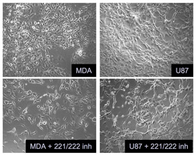
b) Continuous Cell Lines
They are derived from primary cell lines and are transformed/cancerous cells. They can exist in either polyploid form or multiploid form and theoretically can be sub-cultured indefinitely. It is a method of choice of cultivating viruses, where possible.
Photo micrograph of continuous cell lines (cancerous cells present)
Okay firstly, how do we study viruses? These various methods are categorised into 2 groups.
1. For isolation and cultivation of viruses.
Animals/ plants
Embryonated eggs
Tissue culture
2. For detection, identification and diagnosis of viruses.
Tissue culture methods
Physical methods
Serological methods
Immunological methods
Others and Molecular Biology
Each of the following methods will be elaborated in our next posts. Want to find out more? Do read up our upcoming posts so you will be updated on this indeed interesting microbiology topic! (:
Welcome to our Microbiology B Blog ! This blog is created by Chloe, Fatin, Siew Hoon and Shirlene. We are from MB0803 and our assignment will be on the methods of study of viruses.
HELLO THERE! You are reading the microbiology blog done by Shirlene Poon Jia Wen , Chloe Wee Luyi , Sim Siew Hoon and Fatin Nadhirah of MB0803! Hope you enjoy learning about the methods of study of viruses. We made it as interesting as possible. HAHA. Have fun reading and understanding (:


 Reference:
  Reference :
 Reference:
 Reference :

 Reference :
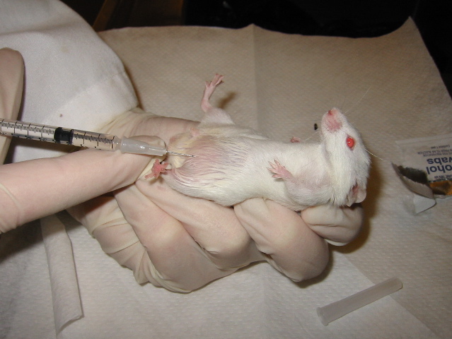