Monday, February 2, 2009

Serological / Immunological Methods

Let's continue with the alternative methods, which are serological/ immunological methods. These methods consist of :

  • Haemagglutination (HA)
  • Haemagglutination Inhibition (HI)
  • Virus neutralisation
  • Complement fixation
  • Immunostaining
  • Immunoprecipitation/ Immunoblot
  • ELISA

Unfortunately yes, there are many methods to know about. Let's go through one by one slowly..

1. Haemagglutination

Haemagglutination is visible macroscopically and is the basis of haemagglutination tests to detect the presence of viral particles. The test does not discriminate between viral particles that are infectious and particles that are degraded and no longer able to infect cells. Both can cause the agglutination of red blood cells.

-Influenza and other viruses
-Two spike proteins: NEURAMINIDASE , HAEMAGGLUTININ ( Binds specifically to red blood cells)

Steps to haemagglutination:
1. Dispense diluent.
2. Add red blood cells and mix by gently shaking.
3. Allow the red blood cells to settle and observe the pattern.
4. Observe if the cells have a normal settling pattern and there is no auto-agglutination. This will be a distinct button of cells in the micro test and an even suspension with no signs of clumping in the rapid test.


2. Haemagglutinin Inhibition

Serologic tests in which a known quantity of antigen is added to the serum prior to the addition of a red cell suspension. Reaction result is expressed as the smallest amount of antigen which causes complete inhibition of haemagglutination.
It is possible to carry out rapid haemagglutination tests and haemagglutination-inhibition tests on a plate, just as for the bacterial agglutination tests described above. However HA and HI are generally only used in this way to confirm the presence and identity of a haemagglutinating antigen.
  • HA conducted in the presence of antibody
  • Neutralisation of virus inhibits agglutination


3. Virus Neutralisation

Antibody neutralisation prevents/ lowers virus infectivity.

4. Complement fixation

The complement fixation test is an immunological medical test that can be used to detect the presence of either specific antibody or specific antigen in a patient's serum. It was widely used to diagnose infections, particularly with microbes that are not easily detected by culture methods, and in rheumatic diseases. However, in clinical diagnostics labs it has been largely superseded by other serological methods such as ELISA and by DNA-based methods of pathogen detection, particularly PCR.
  • Meditated by antibody
  • Antibody binds to antigen

  • Complement cascade of molecules in blood serum initiated, causing lysis of infected cell or pathogen


5. Immunostaining

- Immunoflourescence

-Immuno-gold EM

Immunoflourscence: A laboratory technique to identify specific antibodies or antigens. Antibody identification is usually performed on blood (serum).

  • Antibody tagged with flourescent dye
  • Antibody attached specifically to antigen
  • View specimen under exciting light
  • Flourscence microscope


Immunogold Electron Microscopy :
  • Same principle as immunoflourscence
  • Gold particles attached to antibodies (nanometer size particles)

  • Viewed under EM to localise specific proteins or antigens


6. Immunoprecipitation/Immunoblot

Immunoprecipitation:

- Antigen radioactivity labelled
- Reaction with anitibody
- Isolate anitibody –anitigen complex
- Run through SDS-PAGE
- Detect through x ray film


Immunoblot:

- Western blot analysis
- Whole protein sample run through SDS-PAGE
- Transfer to nitrocellulose membrane
- Antibody attaches specifically to one protein
- Antibody labelled with sensitive indicator
- Colour reaction with streptavidin


7. (LASTLY.) ELISA

ELISA is a widely-used method for measuring the concentration of a particular molecule (e.g., a hormone or drug) in a fluid such as serum or urine. It is also known as enzyme immunoassay or EIA.
The molecule is detected by antibodies that have been made against it; that is, for which it is the antigen. Monoclonal antibodies are often used.

The test requires:
- the antibodies fixed to a solid surface, such as the inner surface of a test tube;
- a preparation of the same antibodies coupled to an enzyme. This is one (e.g., β-galactosidase) that produces a colored product from a colorless substrate.

Performing the Test:

1. The tubes are filled with the antigen solution (e.g., urine) to be assayed. Any antigen molecules present bind to the immobilized antibody molecules.

2. The antibody-enzyme conjugate is added to the reaction mixture. The antibody part of the conjugate binds to any antigen molecules that were bound previously, creating an antibody-antigen-antibody "sandwich".

3. After washing away any unbound conjugate, the substrate solution is added.

4. After a set interval, the reaction is stopped (e.g., by adding 1 N NaOH) and the concentration of colored product formed is measured in a spectrophotometer. The intensity of color is proportional to the concentration of bound antigen.

ELISA can also be adapted to measure the concentration of antibodies. In this case,

1. The wells are coated with the appropriate antigen.

2. The solution (e.g., serum) containing antibodies is added.

3. After they have had time to bind to the immobilized antigen,

4. an enzyme-conjugated anti-immunoglobulin is added, consisting of

- an antibody against the antibodies being tested for. For example, if human anti-HIV antibodies are being assayed, then antibodies (raised in a goat or rabbit against human immunoglobulins) are conjugated to
- the enzyme.

5. After washing away unreacted reagent, the substrate is added.

6. The intensity of the color produced is proportional to the amount of enzyme-labeled antibodies bound (and thus to the concentration of the antibodies being assayed).

Literally hundreds of ELISA kits are manufactured for
- research
- human and veterinary diagnosis

Some examples:
-screening donated blood for evidence of viral contamination
-measuring hormone levels
-detecting infections

That's all for the serological/immunological methods! It's a very long list but we hope you all understand the methods better with our explanations and diagrams.

The very last method used for detection will be up in the next post!

stay tuned.

:DD

Sunday, February 1, 2009

Physical Methods

There are three general types of physical methods for detection, identification and diagnosis.


These methods are :
1) X-ray Crystallography

2) Electron Microscopy (EM)
- Transmission EM
- Scanning EM
- STEM

3) Ultracentrifugation
- Purification
Now, I shall talk about the first physical method, which is X-ray Crystallography

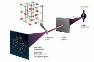
X-RAY CRYSTALLOGRAPHY


Source : http://www.district87.org/staff/sutterm/X-Ray/X-Ray/Intro%20to%20Xray%20Crys.htm

What is X -Ray Crystallography?

It is the study of crystal structures by applying the x-ray diffraction techniques.

Basically, when an Xray beam bombards a crystalline lattice in a specific type of orientation, the X-ray beam is divided in a definite way that was characterised by the lattice's atomic structure. This phenomenon is known as X-Ray diffraction and occurs when the wavelength of X-rays and the interatomic distances within the lattice have the exact magnitude order.

There are three main steps in X-ray crystallography.

1st Step (Often the most challenging step) :

Obtain an adequate crystal of material under study. The crystal should be large enough, maybe larger than 100 micrometres in all dimensions, pure in composition and has a regular structure, with no significant internal imperfections (cracks/twinning).

Conversely, a small or irregular crystal will give fewer and less accurate data, whereby it may be impossible to determine the atomic arrangement.

2nd step:
The crystal is placed in an intense beam of X-rays, usually monochromatic X-rays, that produces the regular pattern of reflections. Previous reflections disappear and new ones appear as the crystal is gradually rotated. The intensity of each spot is recorded at each single orientation of the crystal. In this case, several data sets may have to be collected with each set covering slightly more than half a complete rotation of the crystal and usually containing ten thousands of reflection intensities.

3rd step :

These collected data are compiled computationally with complementary chemical information in order to create and refine a model of the atomic arrangement within the crystal itself. The refined and final model of the atomic arrangement (Crystal Structure) is usually stored in a public database.

ELECTRON MICROSCOPY

What is an electron microscope?
An electron microscope is a type of microscope that uses electrons to illuminate a specimen and create an enlarged image. Electron microscopes have much greater resolving power than light microscopes and can obtain much higher magnifications.

ADD EM FROM PICTURES.

Transmission electron microscope:
The original form of electron microscope, the transmission electron microscope (TEM) uses a high voltage electron beam to create an image. The electrons are emitted by an electron gun, commonly fitted with a tungsten filament cathode as the electron source.


Scanning electron microscope:
Unlike the TEM, where electrons of the high voltage beam carry the image of the specimen, the electron beam of the Scanning Electron Microscope (SEM) does not at any time carry a complete image of the specimen. The SEM produces images by probing the specimen with a focused electron beam that is scanned across a rectangular area of the specimen.


Reflection electron microscope:
In the Reflection Electron Microscope (REM) as in the TEM, an electron beam is incident on a surface, but instead of using the transmission (TEM) or secondary electrons (SEM), the reflected beam of elastically scattered electrons is detected. This technique is typically coupled with Reflection High Energy Electron Diffraction and Reflection high-energy loss spectrum (RHELS).


Scanning Transmission Electron Microscope (STEM):
The STEM rasters a focused incident probe across a specimen that (as with the TEM) has been thinned to facilitate detection of electrons scattered through the specimen. The high resolution of the TEM is thus possible in STEM. The focusing action (and aberrations) occur before the electrons hit the specimen in the STEM, but afterward in the TEM.

ULTRACENTRIFUGATION

What is ultracentrifugation?

Ultracentrifugation is a centrifuge optimized for spinning a rotor at very high speeds, capable of generating acceleration as high as 1,000,000 g (9,800 km/s²). There are two kinds of ultracentrifuges, the preparative and the analytical ultracentrifuge.

PUT ULTRACENTRIFUDGE 1 IN PICTURES.

Source : http://www.mmb.usyd.edu.au/facilities/images/analytical_ultracentrifugation300x211.jpg


Next Up : Cytopathic Effect (CPE)

Cytopathic Effect (CPE) is the second tissue culture method for detection of viruses. Yes, bear with me. This method is not as time-consuming as plaque assay. What is it exactly? Let's find out..

Cytopathic effect (CPE) refers to degenerative changes in cells (especially in tissue culture) associated with the multiplication of certain viruses. When in tissue culture, the spread of virus is restricted by an overlay of agar (or other suitable substance) and thus the cytopathic effect may lead to formation of plaque.

Changes include:

  • Altered Shape
  • Detachment from substrate
  • Lysis
  • Membrane fusion
  • Altered membrane permeability
  • Inclusion bodies
  • Apoptosis

(a) Host cell damage:

(i) Cytopathic effects are damage to infected host cells caused by infecting viruses .

(ii) Basically, viral infection can lead to cell abnormalities (biochemical and morphological) and/or cell death.


(b) Not exactly lysis:

(i) The damage defining cytopathic effects or death (cytocidal effects) is unlike that caused by lytic viruses upon the release of progeny virus .

(ii) That is, lysis is a very purposeful killing of the host cell.

(iii) Cytopathic effects instead are a consequence of the virus' metabolic needs and those of the host cell simply not completely corresponding.


Pictures of CPE:


That's all for CPE. Next post will be on the other detection methods. Such as the physical and serological methods. Keep on reading to be updated!

:DD

Saturday, January 31, 2009

Plaque Assay. (not ESSAY.)

Let's move on to the detection, identification and diagnosis of viruses.
okay one of the tissue culture methods detection is PLAQUE ASSAY. What is it and how does it work? Let's find out together, shall we?

Plaque Assay:

A method of quantifying the number of infectious units by inoculating serial dilutions of a viral suspension on a cell culture monolayer, overlaying with a medium containing agarose and after several days incubation, counting the number of plaques formed; recorded as plaque forming units/ml. These plaques can sometimes be detected visually using colony counters, in much the same way as bacterial colonies are counted. This technique requires counting the number of plaques formed by a virus sample, from which the actual virus concentration can be determined.

Features:
  • Very time-consuming
  • Very simple method
  • Only works for viruses that infect monolayer cells
  • Only works for viruses that causes cell lysis
  • Uses principle of one virus on the monolayer produces one plaque


To know the steps to do Plaque Assay, please watch this video :


Is it more clearer now? We hope you understood plaque assay better. Stay tuned again! We will be updating on the second method which is Cytopathic Effect (CPE).
till then.
:D
-fatin(:

I-S-O-L-A-T-I-O-N...Not your usual "Lemon Tree" tune.

The title makes no sense, I know. So, why don't we continue with something more logical? =)

Now, I shall explain the methods used for isolation and cultivation :

1) Animals/Eggs

This method was the first method used for virus cultivation purposes. However, this method is inconvenient and extra attention and care is needed when handling animals. Mainly replaced cell culture except for two points :

a) The virus has no known host in vitro
For instance, Hepatitis C virus in chimpanzees and Influenza virus in chick embryo.


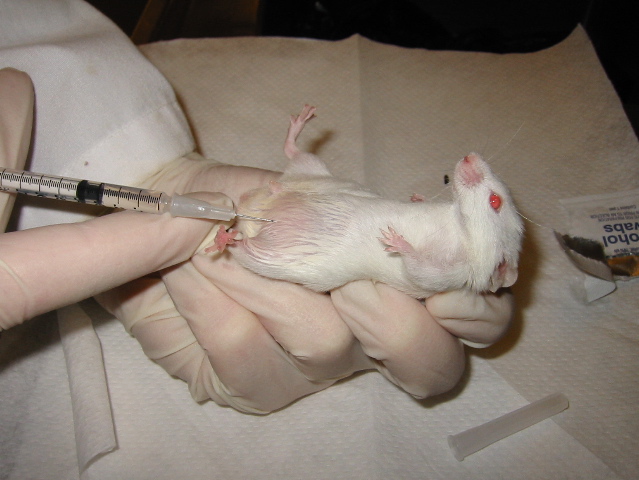
Diagram of mouse infection :

Here's a video about a successful intravenous dye injection in mice. The same principle applies in virus injection in mice :

Diagram of growth of viruses in embryonated eggs :
Reference: http://www.cmbi.bjmu.edu.cn/

b) Study of viral pathogenesis in a whole host
For example, polio studies in chimpanzees.
Pathogenesis of polio virus

Humans are the only natural host of polio virus. Polio viruses have a tropism for the epithelial cells lining the alimentary tract and for cells of the central nervous system. They attach to a specific receptor on these cells, which in humans is encoded by a gene on chromosome 19. Polio virus infection is quite common in nonimmunized individuals, but only about 1 percent of these cases progress to the paralytic form of the disease.
The histocompatibility antigens HLA-3 and HLA-7 are believed to be highly associated with an increased risk of paralysis. Primary replication of polio virus takes place in the oropharyngeal and intestinal mucosa (the alimentary phase).

From here, the virus spreads to the tonsils and Peyer's patches of the ileum and to deep cervical and mesenteric nodes, where it multiplies abundantly (the lymphatic phase). Subsequently, the virus is carried by the bloodstream to various internal organs and regional lymph nodes (the viremic phase).
In most cases, no further virus spread occurs, and there is asymptomatic or mild febrile undifferentiated illness, such as fever, malaise, headache, nausea, gastrointestinal disturbances, and sore throat, or combination of these.

Illustration of a Poliovirus


Diagram of an African Green Monkey Cell

Reference : http://www.nature.com/

2) Plants
Plants are used in the study of Tobacco mosaic virus and this is based on the virus plaques on the leaves of plants to determine the virus numbers.
A photomicrograph of Tobacco Mosaic Virus (TMV)

Reference : http://www.erec.ifas.ufl.edu/
The Tobacco mosaic virus (TMV) is an RNA virus that infects plants, tobacco in particular, showing characteristic patterns like mottling and discoloration on the leaves, hence the name. It was the first virus to be discovered.

Picture of a leaf used in the study of TMV (Gradual changes shown in A and B)
Reference : http://www.apsnet.org/
Fig. 1. Tobacco mosaic virus (TMV). (A) Systemic infections of Nicotiana tabacum cv. Turk plants showing TMV-associated mosaic. (B) Necrotic local lesions on N. tabacum Glurk leaf, demonstrating Holmes’ N-gene resistance following inoculation with TMV.


3) Tissue Culture

In cell tissue culture, the cells are grown in vitro, which means they are grown in test tubes.

Primary cell cultures typically will have a finite life span in culture whereas continuous cell lines are, by definition, abnormal and are often transformed cell lines.

a) Primary Cell Cultures

b) Continuous Cell Lines

They are derived from primary cell lines and are transformed/cancerous cells. They can exist in either polyploid form or multiploid form and theoretically can be sub-cultured indefinitely. It is a method of choice of cultivating viruses, where possible.

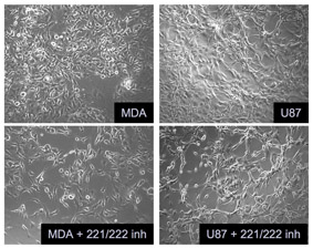
Photo micrograph of continuous cell lines (cancerous cells present)

Reference : http://research.nki.nl/agamilab/images/p27.jpg


TO BE CONTINUED (Trying to find more information and...pictures!)
Shir

THE METHODS OF STUDY OF VIROLOGY!

Okay firstly, how do we study viruses? These various methods are categorised into 2 groups.

1. For isolation and cultivation of viruses.
  • Animals/ plants
  • Embryonated eggs
  • Tissue culture

2. For detection, identification and diagnosis of viruses.

  • Tissue culture methods
  • Physical methods
  • Serological methods
  • Immunological methods
  • Others and Molecular Biology

Each of the following methods will be elaborated in our next posts. Want to find out more? Do read up our upcoming posts so you will be updated on this indeed interesting microbiology topic! (:

stay tuned!

:D

Introduction

Welcome to our Microbiology B Blog ! This blog is created by Chloe, Fatin, Siew Hoon and Shirlene. We are from MB0803 and our assignment will be on the methods of study of viruses.